


Dear Editor,
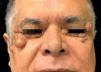
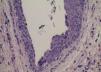
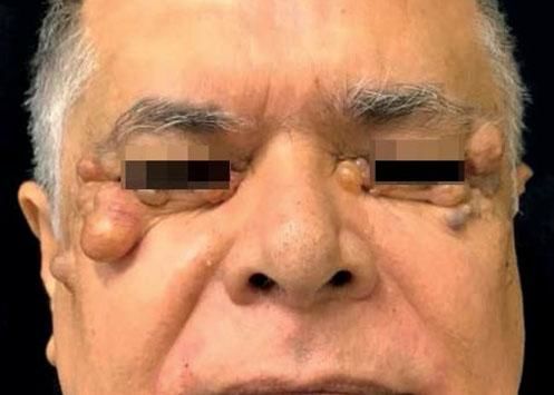
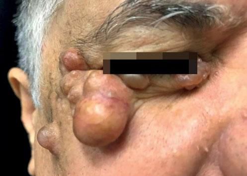
Hidrocystoma is a benign tumor originating from the apocrine gland, being an adenoma of this gland, unlike the eccrine hidrocystoma that results from a ductal dilatation by retention of secretions. It presents as translucent, round, small, painless vesicles with fluid content in their interior.1 Traditionally, they are divided into solitaries (Smith type) and multiples (Robinson type).2 Clinical differences that help in their diagnosis are: apocrine is usually solitary, larger, with a bluish color, although translucent and generally located on the face, especially on the lower palpebral region (cysts of Moll's glands) near the cilia and lacrimal drainage pathway; eccrine may be solitary or multiple, may increase with heat and decrease in the cold, translucent or opaque, with a more frequent location, on the lower eyelids but above the palpebral skin.3 They are also found on other regions such as ear, trunk, scalp, and upper limbs. Generally, they occur in adults, especially females, after the 4th decade of life. The case reported is of a 62-year-old male, white patient, who sought a dermatology clinic with the following complaint: "lumps on the face for more than five years." At the dermatological examination, there were skin-colored papules and nodules on the periocular region, with a shiny surface, translucent appearance and rare telangiectasias (Figure 1 and 2). An excisional biopsy of a nodule was performed and the histopathological examination showed a cystic lesion with a thin layer of cuboidal epithelial cells with apocrine features and amorphous liquid content, with no signs of malignancy (Figure 3). Surgical excision of the lesions was scheduled, but patient did not return to the service.
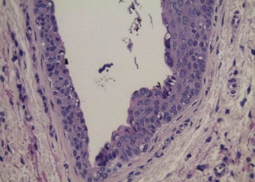
Its pathogenesis seems to result from obstruction of the sweat duct just above the glandular groove (deep dermis) due to an inflammatory process or trauma. The diagnosis is initially clinical, followed by histological confirmation. Histologically, apocrine hidrocystomas are unilocular or multilocular dermal cysts with one or more layers of epithelial cells showing bulbous protrusions and luminal secretion by decapitation.4 It may also have papilliferous projections, being covered by two layers of secretory cells. The inner cells are columnar and show eosinophilic cytoplasm with typical bulboapical expansions.
The main clinical differential diagnoses include: molluscum contagiosum, nodulocystic basal cell carcinoma, hidradenoma, nevocytic nevus, blue nevus, syringoma, hordeolum, chalazion, epidermal cyst. Treatment can be done through surgical excision, shaving and electrocoagulation, cryosurgery or even CO2 laser, motivated by the unsightly aspect of the lesions.5
AcknowledgementsWe would like to thank the whole team for the commitment in dealing with the case.
