Journal Information
Vol. 94. Issue 4.
Pages 497-498 (01 July 2019)
Share
Share
Download PDF
More article options
Vol. 94. Issue 4.
Pages 497-498 (01 July 2019)
Open Access
Nodular amelanotic melanoma*
Visits
3723
Thadeu Santos Silva1, Luciana Rebouças de Araujo2, Gustavo Baptista de Almeida Faro1, Geise Rezende Paiva3
1 Dermatology Outpatient Clinic, Hospital Universitário Professor Edgard Santos, Universidade Federal da Bahia, Salvador, BA, Brazil.
2 Dermatology Outpatient Clinic, Hospital Santa Izabel, Escola Bahiana de Medicina e Saúde Pública, Salvador, BA, Brazil.
3 Anatomical Pathology Laboratory, Hospital Santa Izabel, Escola Bahiana de Medicina e Saúde Pública, Salvador, BA, Brazil.
This item has received

Under a Creative Commons license
Article information
Full Text
Bibliography
Download PDF
Statistics
Figures (3)
Show moreShow less
Full Text

Dear Editor,

Amelanotic melanomas are characterized by absence of pigment.1 They are an infrequent variant of classically described clinical and pathological subtypes, estimated occur in between 2 and 8% of all cases.1,2 Amelanotic melanomas may present as erythematous maculae on sun-exposed areas, with epidermal alterations, normochromic dermal plaques, without epidermal changes or papulonodular lesions.1 The most common form is the nodular variant, but any other clinicopathological form can be found.2 Dermoscopy may serve as a valuable tool for elucidating the suspicion of this entity.2 Although structures that suggest the presence of melanin are absent, it is possible to visualize vascular patterns and residual pigmentation that are not visible to the naked eye.3 The predominant vascular pattern in melanocytic and hypomelanotic melanomas depends on tumor thickness.2 Diagnosis is also hampered by the absence of well-established criteria.4 In addition, they do not differ from pigmented melanomas in terms of prognosis or treatment.1

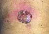
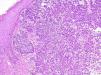
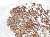
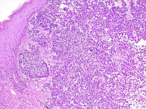
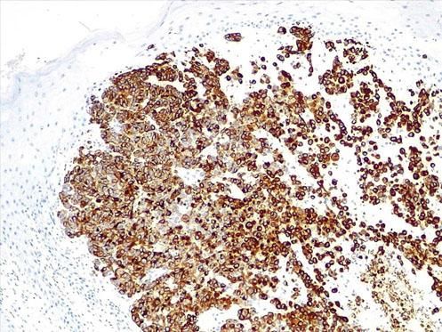
Male patient, 60 years old, with diagnosis of schizophrenia, presented a tumoral lesion approximately 4cm in diameter, pedunculated, in the lumbar region, with a peritumoral erythematous area (Figure 1). The lesion was excised with 2mm margins. Anatomopa-thological examination evidenced an extensive area of ulceration, covered by fibrinous exudate. The dermis showed a malignant neoplasm composed of large epithelioid and fusiform cells, forming cohesive and extensively infiltrative blocks and areas of necrosis. The cells exhibited intense hyperchromasia and pleomorphism, with numerous typical and atypical mitotic figures, evident nucleoli and eosinophils, and presence of brownish pigment (Figure 2). Immunohistochemistry was positive for Melan A, S100, SOX10, and HMB45, confirming the diagnosis of malignant melanoma, nodular type, with Breslow thickness 1.5cm and mitotic index of 15 mitoses/mm2 (Figure 3). Lymphatic, blood, and perineural invasion were not detected. Surgical margins were free, 2.6 mm away from the neoplasia.

Figure 1.

Tumoral lesion approximately 4cm in diameter, located on the lower back, with surrounding erythema

(0.03MB).
Figure 2.

Histopathology: Confluence of nests of large, atypical melanocytes. The cells are irregular in size, shape, and spacing. Many have hyperchromatic and pleomorphic nuclei and pigmented cytoplasm (Hematoxylin & eosin, x100)

(0.07MB).
Figure 3.

Immunohistochemistry: expression of HMB45 (HMB45, x100)

(0.07MB).

Pgmentation is one of the guiding criteria for the diagnosis of melanocytic lesions, and lack of pigmentation often makes the diagnosis of amelanotic melanoma challenging.1,2 In such cases, dermoscopy is important for the evaluation of morphological types of vascular structures.3 The vascular pattern may not be sufficient, and it is essential to associate the clinical findings and patient’s history.3 The definitive diagnosis can only be established by histopathology, and immunohistochemistry is often necessary.2,4 The present report highlights the importance of histopathology and immunohistochemistry for diagnostic elucidation. The lack of defined criteria can delay the suspicion and diagnosis, fundamental to prognosis in this highly aggressive and potentially lethal entity.4

Acknowledgments

The authors wish to thank Dr. Geise Rezende Paiva for her collaboration and dedication in preparing the histopathology and immunohistochemistry images.

References
[1.]
Thomas NE, Kricker A, Waxweiler WT, Dillon PM, Busman KJ, From L, et al.
Genes, Environment, and Melanoma (GEM) Study Group..
Comparison of clinicopathologic features and survival of histopathologically amelanotic and pigmented melanomas: a population-based study. JAMA Dermatol., 150 (2014), pp. 1306-1314
[2.]
Cheung WL, Patel RR, Leonard A, Firoz B, Meehan SA..
Amelanotic melanoma: a detailed morphologic analysis with clinicopathologic correlation of 75 cases..
J Cutan Pathol., 39 (2012), pp. 33-39
[3.]
Jaimes N, Braun RP, Thomas L, Marghoob AA..
Clinical and dermoscopic characteristics of amelanotic melanomas that are not of the nodular subtype..
J Eur Acad Dermatol Venereol., 26 (2012), pp. 591-596
[4.]
Mendes MS, Costa MC, Gomes CM, de Araújo LC, Takano GH..
Amelanotic metastatic cutaneous melanoma..
An Bras Dermatol., 88 (2013), pp. 989-991

Study conducted at the Pathology Service, Escola Bahiana de Medicina e Saude Publica, Salvador, BA, Brazil.

Financial support: None.

Conflict of interest: None.

Copyright © 2019. Anais Brasileiros de Dermatologia
Idiomas
Anais Brasileiros de Dermatologia
Article options
Tools
en pt
Cookies policy Política de cookies
To improve our services and products, we use "cookies" (own or third parties authorized) to show advertising related to client preferences through the analyses of navigation customer behavior. Continuing navigation will be considered as acceptance of this use. You can change the settings or obtain more information by clicking here. Utilizamos cookies próprios e de terceiros para melhorar nossos serviços e mostrar publicidade relacionada às suas preferências, analisando seus hábitos de navegação. Se continuar a navegar, consideramos que aceita o seu uso. Você pode alterar a configuração ou obter mais informações aqui.