


Onychocytic matricoma is a newly described tumor of the nail matrix. Clinically, it presents with localized thickening of the nail plate and melanonychia. Histologically, it represents a benign acanthoma of onychocytes. There are 8 cases reported in the literature. A 12-year-old girl presented with localized melanonychia and concurrent thickening of the nail plate restricted to the area of pigmentation affecting the right thumb, with no history of trauma or pain. We report a case of this rare tumor occurring in late childhood and provide a comprehensive review of its clinical presentation and differential diagnosis. Both clinicians and dermatopathologists should be aware of the presentation of onychocytic matricoma and include it in their scope of diagnosis of longitudinal nail bands.
Matrical tumors are a diverse group of neoplasms composed of cells with matrical differentiation. Onychomatricoma was the only matrical tumor identified in the nail unit until Perrin et al. described the onychocytic matricoma in 2012.2,3 This tumor typically presents as a localized thickening of the nail plate that may display concurrent melanonychia and represents a benign acanthoma of on-ychocytes. We report a case of onychocytic matricoma occurring in late childhood and provide a comprehensive review of its clinical presentation and differential diagnosis.
CASE REPORTA 12-year-old female noticed a dark line under the nail 3 months prior to consultation. No history of traumatic injury, pain, or any other symptom was reported. Physical examination showed a 2-mm-wide black longitudinal streak starting at the distal lunula with localized nail plate thickening on the right thumb (Figure 1). Dermoscopy revealed a sharply delimitated longitudinal melanonychia seen on the dorsal surface of the nail plate and localized thickening of the nail plate with subungual hyperkeratosis at the hyponychium.
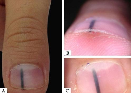
A - Physical examination of 2-mm-wide black longitudinal streak starting at the distal surface of the right thumb. B - Distal dermoscopy highlights a localized thickening of the nail plate with subungual hyperkeratosis at the hyponychium. C - Dorsal plate dermoscopy shows a sharply delimitated longitudinal melanonychia on the dorsal surface of the nail plate
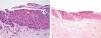
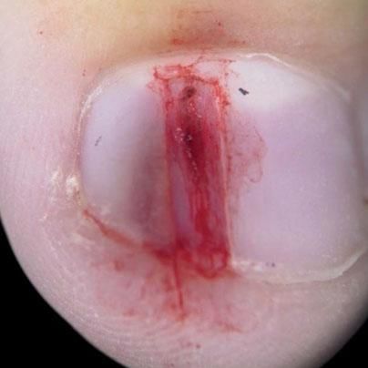
The risks of biopsy and excision were discussed with the family, who opted for the complete removal of the lesion and partial removal of the nail plate (Figure 2). Histopathological analysis revealed moderate acanthosis of the nail matrix, with proliferation of matrical onychocytes with small monomorphic nuclei with condensed chromatin and scant cytoplasm. Serial sections showed a single area of keratinization simulating a squamous eddy. Fontana Masson stain revealed melanin in the cytoplasm of matrical onychocytes and sparse melanocytes in the dendrites (Figure 3). Clinical-pathological correlation provided for the diagnosis of onychocytic matricoma, a pigmented acanthotic variant.
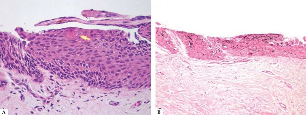
Histopathological analysis revealed: A - Hematoxylin & eosin (x200) moderate acanthosis of the nail matrix, with proliferation of matrical onychocytes with small monomorphic nuclei with condensed chromatin and scant cytoplasm. Arrow: area of keratinization simulating a squamous eddy. B - Fontana Masson (x100): melanin within the cytoplasm of matrical onychocytes and sparse melanocytes in the dendrites
Onychocytic matricoma is a rare tumor of the nail matrix first described in 2012. Only 8 cases have been reported to date. Table 1 summarizes the clinical, epidemiological, and histopathological features. The tumor only affects the fingernails and may present as a localized longitudinal melanonychia, often associated with thickening of the nail plate that can simulate a foreign body, nevus, or even melanoma.1,2,3 There is one case report of hypopigmented type that was characterized by a band of xanthonychia obscuring the lunula, also presenting splinter hemorrhages.4 According to previous case reports, the patient may notice the tumor 3 months to 4 years before seeing a dermatologist, and age at onset can vary from 12 to 80 years.1-4
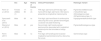
Summary of the clinical, epidemiological, and histopathological features of published case reports/case series
| Sex | Age | History of trauma | Clinical Presentation | Histologic Variant | |
|---|---|---|---|---|---|
| Perrin et al. (2012) | Female (4)Male (1) | 17-80 | no (3) | Fifth digit, right hand (2)Third digit, right hand (2)First digit, right hand (1)All lesions presented as longitudinal melanonychia | Pigmented (4)Melanocytic (1)Acanthotic type (2)Keratogenous type (2)Papillomatous type (1) |
| Spaccarelli et al. (2013) | Male | 69 | no | First digit, right hand Band of xanthonychia obscuring the lunula, splinter hemorrhages. Localized nail plate thickening | HypopigmentedAcanthotic type |
| Wannat et al. (2014) | Male | 40 | yes | Second digit, right handLocalized pachyonychia associated with longitudinal melanonychia that simulates a foreign body | PigmentedKeratogenous type |
| Perrin (2017) | Male | 50 | no | First digit, left handAcquired localized longitudinal pachymelanonychia, obscuring the lunula | Germinotropic type |
Histologically, onychocytic matricoma consists of a basal compartment with a varying admixture of prekeratogenous and keratogenous cells. Two particular features are the concentrically arranged nests of these cells and the endokeratinization of the tumor’s deep portion. It can be divided into four variants: acanthotic type, when distinct acanthosis is present; acanthotic and papillomatous type, characterized by acanthosis with an increased proportion of basaloid cells, the presence of spheres of prekeratogenous and keratogenous zone cells located in the basaloid compartment, distal superficial epithelial papillae, and a thick nail plate with multiple cavities; keratogenous type, which has a prominent keratogenous zone in the presence of a delayed maturation pattern; and the germinotropic variant showing prominent basaloid compartment with peripheral palisading, mimicking the appearance of a basal cell carcinoma. Another classification that is based on pigmentation can also be used: pigmented, melanocytic, and nonpigmented.1
Differential diagnosis should include foreign body, nevus, onychomatricoma, onychopapilloma, and onychocytic carcinoma. Moreover, irritated seborrheic keratosis is among the histological differentials.1-4
Onychomatricoma, onychocytic matricoma, and onychocytic carcinoma are the only matrical tumors that arise from the nail matrix. Clinically, onychomatricoma presents as a yellow longitudinal band of variable width that may be hidden under melanonychia. Splinter hemorrhages may also be seen. The microscopy is characterized by nail bed papillomatosis within a fibrous and cellular stroma, whereas the papillomatous variant of onychocytic matricoma (the main differential) has superficial epithelial digitations that lack a connective tissue core.5 Onychocytic carcinoma presents as an acquired localized (monodactylous) longitudinal pachyonychia. Histologically, this in situ malignant epithelial tumor exhibits cytological features of malignancy, including marked nuclear pleomorphism, prominent nucleoli, and frequent mitotic figures, many of which are abnormal.2,6
Onychopapilloma presents as a longitudinal yellow-white band with occasional splinter hemorrhages that may be associated with melanonychia and erythronychia. It differs histologically from onychocytic matricoma because it is a tumor of the nail bed instead of nail matrix and is characterized by acanthosis in the presence of nail bed papillomatosis.7
Subungual seborrheic keratosis is a yellowish-white longitudinal band with splinter hemorrhages and subungual localized hyperkeratosis. However, a helpful additional feature is the presence of white, round-shaped “milia-like” cysts that are observed through the translucent nail plate.8 The irritated subtype is the entity that can mimic onychocytic matricoma histologically. However, they can be differentiated by the type of cornification, the latter mainly composed of nail plate cells and irritated seborrheic keratosis by squamous eddies with apoptotic cells.9
We report herein a case of onychocytic matricoma in a 12-year-old female that presented clinically as a foreign body. The authors call the attention of clinicians and dermatopathologists to the existence of onychocytic matricoma in order to add this entity to the differential diagnosis of longitudinal nail bands.
