Journal Information
Vol. 94. Issue 4.
Pages 484-485 (01 July 2019)
Share
Share
Download PDF
More article options
Vol. 94. Issue 4.
Pages 484-485 (01 July 2019)
Open Access
P-wave dispersion in vitiligo patients*
Visits
3590
Soheila Nasiri1, Mehdi Pishgahi2, Soma Ahmadi1, Sahar Dadkhahfar1
1 Skin Research Center, Shohada-e Tajrish Hospital, Shahid Beheshti University of Medical Sciences, Tehran, Iran.
2 Department of Cardiology, Shohada-e Tajrish Hospital, Shahid Beheshti University of Medical Sciences, Tehran, Iran.
This item has received

Under a Creative Commons license
Article information
Full Text
Bibliography
Download PDF
Statistics
Tables (1)
Table 1. P-max P-min, and P-wave dispersion (PWD) across the groups
Full Text

Dear Editor,

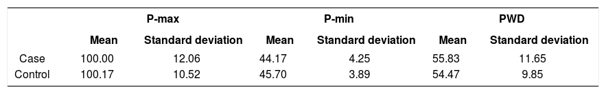
Vitiligo is an acquired depigmentation disorder of unknown origin.1 Melanocytes are found in the heart valves and septum, the atrium, and the vessels, with possible role in abnormal signals and atrial arrhythmia.2 P-wave dispersion (PWD) is the difference between P-max and P-min in 12-lead electrocardiography.3 Assessment of possible risk is useful, especially regarding the accessibility of electrocardiography in all health care centers and the possible association between some electrocardiogram (ECG) alterations and the presence of arrhythmia, even in healthy subjects. Hence, the purpose of this study was to determine the PWD in vitiligo patients vs. control subjects. A case-control study was performed in a referral dermatology outpatient clinic in Shohada-e-Tajrish Hospital, affiliated with the Shahid Beheshti University of Medical Sciences, Tehran, Iran, during a one-year period (October 2016 to October 2017). A total of 94 participants were included in this study, i.e., 47 patients with vitiligo and 47 healthy volunteers. The exclusion criteria were history of other diseases, smoking, and obesity. The medical assessment involved natural and Wood’s light examinations consistent with the modified Vitiligo European Task Force form. Based on the Vitiligo European Task Force, a patient’s palm, corresponding to 1% of body surface area, was used to calculate the total vitiligo extension. The P-max and P-min were calculated in all 12 ECG leads and the difference between them was defined as the PWD. Data analysis was performed using SPSS (v 18.0) software, utilizing the chi-squared test and Student’s f-test for independent samples. Statistically significant p-values were those less than 0.05. In each group, 32 patients were female. The mean (standard deviation) age was 34.9 (12.1) and 32.8 (11.3) years in the case and control groups, respectively (p > 0.05). The vitiligo type was vulgaris in 87.2% and acrofacial in 12.8%. The mean involved body area by vitiligo in case group was 30%. The results in Table 1 demonstrate that PWD was alike across the case groups (p > 0.05). The EF, heart rate, and blood pressures had no significant differences (p > 0.05). Baldini ef al. reported that vitiligo has been associated with other rare systemic disorders.4 However, there was no significant association in the present study. Fairfax ef al. found an association between vitiligo, primary hypothyroidism, pernicious anemia, and idiopathic chronic heart block.5 In sum, it may be concluded that PWD in vitiligo patients vs. control subjects is not significantly differed. Hence, there is no screening requirement for cardiovascular disorders in patients with vitiligo.

Table 1.

P-max P-min, and P-wave dispersion (PWD) across the groups

  P-maxP-minPWD
  Mean  Standard deviation  Mean  Standard deviation  Mean  Standard deviation 
Case  100.00  12.06  44.17  4.25  55.83  11.65 
Control  100.17  10.52  45.70  3.89  54.47  9.85 
Acknowledgments

The authors would like to thank the patients participating in this study.

References
[1.]
Parker CN, Finlayson KJ, Shuter P, Edwards HE..
Risk factors for delayed healing in venous leg ulcers: a review of the literature..
Int J Clin Pract., 69 (2015), pp. 967-977
[2.]
Fernandez ML, Upton Z, Shooter GK..
Uric acid and xanthine oxidoreductase in wound healing..
Curr Rheumatol Rep., 16 (2014), pp. 396
[3.]
Fernandez ML, Upton Z, Edwards H, Finlayson K, Shooter GK..
Elevated uric acid correlates with wound severity..
Int Wound J., 9 (2012), pp. 139-149
[4.]
Hoffmeister C, Trevisan G, Rossato MF, de Oliveira SM, Gomez MV, Ferreira J..
Role of TRPV1 in nociception and edema induced by monosodium urate crystals in rats..
Pain., 152 (2011), pp. 1777-1788
[5.]
Burnand KG, Whimster I, Naidoo A, Browse NL..
Pericapillary fibrin in the ulcer-bearing skin of the leg: the cause of lipodermatosclerosis and venous ulceration..
Br Med J (Clin Res Ed)., 285 (1982), pp. 1071-1072

Study conducted at the Department of Cardiac Aspects of Skin Diseases, Skin Research Center, Shohada-e Tajrish Hospital, Shahid Beheshti University of Medical Sciences, Tehran, Iran.

Financial support: None.

Conflict of interest: None.

Copyright © 2019. Anais Brasileiros de Dermatologia
Idiomas
Anais Brasileiros de Dermatologia
Article options
Tools
en pt
Cookies policy Política de cookies
To improve our services and products, we use "cookies" (own or third parties authorized) to show advertising related to client preferences through the analyses of navigation customer behavior. Continuing navigation will be considered as acceptance of this use. You can change the settings or obtain more information by clicking here. Utilizamos cookies próprios e de terceiros para melhorar nossos serviços e mostrar publicidade relacionada às suas preferências, analisando seus hábitos de navegação. Se continuar a navegar, consideramos que aceita o seu uso. Você pode alterar a configuração ou obter mais informações aqui.