


We report the case of an 8-year-old child with subungual exostosis, whose diagnosis was suspected on the basis of dermoscopic findings and subsequently confirmed by X-ray and histopathology.
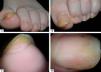
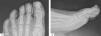
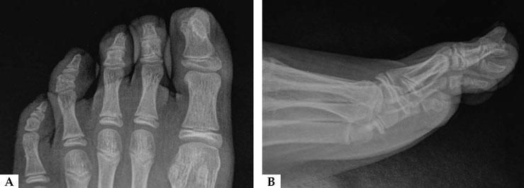
An 8-year-old child presented with a painful subungual nodule on the left hallux, elevating the nail plate (Figures 1A-B). The lesion had appeared 12 months before and had been misdiagnosed as a wart and treated unsuccessfully with cryotherapy. Dermoscopy of the free nail edge showed a combination of vascular ectasia with hyperkeratosis (Figure 1C), while dermoscopy of the nail plate revealed onycholysis (Figure 1D). The combination of these findings was recently reported as repetitive dermoscopic features supporting the diagnosis of subungual exostosis (SE)1, later confirmed by X-ray showing a bony proliferation originating from the distal phalanx of the first toe (Figures 2A–B). Due to increasing pain, the child was referred to pediatric surgery for complete excision of the lesion. Histopathological examination showed microscopic features consistent with the clinical and radiological suspicion of SE.

SE is a benign osteochondral proliferation of the distal phalanx. A recent systematic review of SE showed that half of the cases occur in patients under 18 years of age, although mean age at presentation was 25.7 years.2 Due to these important epidemiological data, it is mandatory to consider SE in the differential diagnosis of subungual proliferation in children, although both benign and malignant tumors are rare in childhood.3 Diagnostic delay is common in SE, since it can be misdiagnosed as viral wart4, with subsequent unnecessary and unsatisfactory treatments.
Our previously published case series showed a mean diagnostic delay of 12 months.1 To avoid diagnostic delay, it should be kept in mind that the bone overgrowth often elevates the nail plate, provoking pain in the patient. Meanwhile, dermoscopy can assist the diagnosis when clinical suspicion of SE arises, subsequently leading to X-ray examination. Members of our group (VP, TR, GA) recently observed the following key dermoscopic features of SE: vascular ectasia as the most common finding (70%), followed by hyperkeratosis (60%), onycholysis (40%), and ulceration (30%)1. In the current case, 3 of these 4 dermoscopic characteristics were identified; importantly, however, dermoscopic findings result from the combination of two acquisitions: nail-plate and free-edge dermoscopy. The previous paper did not specifically mention pediatric patients, but half the patients were under 18 years of age. This new pediatric case corroborates the previous findings, suggesting that careful onychoscopy of both the nail plate and free edge can be highly useful in cases of suspected SE in children. The most common differential diagnosis, viral wart, usually presents a different dermoscopic image, characterized by typical rough surface and dilated capillaries of the papillary dermis, generally visible as hemorrhagic dotted vessels.5
SE is not such a rare benign tumor in children, and for prompt detection, both dermatologists and pediatricians should be alert to the existence of this clinical condition. Dermoscopy orients the differentiation from other nail conditions and the correct treatment choice for the patient.
