array:24 [
  "pii" => "S0365059620300799"
  "issn" => "03650596"
  "doi" => "10.1016/j.abd.2019.07.012"
  "estado" => "S300"
  "fechaPublicacion" => "2020-05-01"
  "aid" => "168"
  "copyright" => "Sociedade Brasileira de Dermatologia"
  "copyrightAnyo" => "2020"
  "documento" => "simple-article"
  "crossmark" => 1
  "licencia" => "http://creativecommons.org/licenses/by/4.0/"
  "subdocumento" => "crp"
  "abierto" => array:3 [
    "ES" => true
    "ES2" => true
    "LATM" => true
  ]
  "gratuito" => true
  "lecturas" => array:1 [
    "total" => 0
  ]
  "Traduccion" => array:1 [
    "pt" => array:19 [
      "pii" => "S2666275220301491"
      "issn" => "26662752"
      "doi" => "10.1016/j.abdp.2019.07.012"
      "estado" => "S300"
      "fechaPublicacion" => "2020-05-01"
      "aid" => "168"
      "copyright" => "Sociedade Brasileira de Dermatologia"
      "documento" => "simple-article"
      "crossmark" => 1
      "licencia" => "http://creativecommons.org/licenses/by/4.0/"
      "subdocumento" => "crp"
      "abierto" => array:3 [
        "ES" => true
        "ES2" => true
        "LATM" => true
      ]
      "gratuito" => true
      "lecturas" => array:1 [
        "total" => 0
      ]
      "pt" => array:12 [
        "idiomaDefecto" => true
        "cabecera" => "<span class="elsevierStyleTextfn">Dermatologia Tropical&#47;Infectoparasit&#225;ria</span>"
        "titulo" => "Pseudomicetoma no couro cabeludo causado por <span class="elsevierStyleItalic">Microsporum canis</span>"
        "tienePdf" => "pt"
        "tieneTextoCompleto" => "pt"
        "tieneResumen" => "pt"
        "paginas" => array:1 [
          0 => array:2 [
            "paginaInicial" => "372"
            "paginaFinal" => "375"
          ]
        ]
        "contieneResumen" => array:1 [
          "pt" => true
        ]
        "contieneTextoCompleto" => array:1 [
          "pt" => true
        ]
        "contienePdf" => array:1 [
          "pt" => true
        ]
        "resumenGrafico" => array:2 [
          "original" => 0
          "multimedia" => array:7 [
            "identificador" => "fig0020"
            "etiqueta" => "Figura 4"
            "tipo" => "MULTIMEDIAFIGURA"
            "mostrarFloat" => true
            "mostrarDisplay" => false
            "figura" => array:1 [
              0 => array:4 [
                "imagen" => "gr4.jpeg"
                "Alto" => 623
                "Ancho" => 1250
                "Tamanyo" => 131052
              ]
            ]
            "descripcion" => array:1 [
              "pt" => "<p id="spar0025" class="elsevierStyleSimplePara elsevierViewall">Aspecto cl&#237;nico ap&#243;s o tratamento&#46;</p>"
            ]
          ]
        ]
        "autores" => array:1 [
          0 => array:2 [
            "autoresLista" => "Ligia Rangel Barboza Ruiz, Clarisse Zaitz, Rute Facchini Lellis, John Verrinder Veasey"
            "autores" => array:4 [
              0 => array:2 [
                "nombre" => "Ligia Rangel Barboza"
                "apellidos" => "Ruiz"
              ]
              1 => array:2 [
                "nombre" => "Clarisse"
                "apellidos" => "Zaitz"
              ]
              2 => array:2 [
                "nombre" => "Rute Facchini"
                "apellidos" => "Lellis"
              ]
              3 => array:2 [
                "nombre" => "John Verrinder"
                "apellidos" => "Veasey"
              ]
            ]
          ]
        ]
      ]
      "idiomaDefecto" => "pt"
      "Traduccion" => array:1 [
        "en" => array:9 [
          "pii" => "S0365059620300799"
          "doi" => "10.1016/j.abd.2019.07.012"
          "estado" => "S300"
          "subdocumento" => ""
          "abierto" => array:3 [
            "ES" => true
            "ES2" => true
            "LATM" => true
          ]
          "gratuito" => true
          "lecturas" => array:1 [
            "total" => 0
          ]
          "idiomaDefecto" => "en"
          "EPUB" => "https://multimedia.elsevier.es/PublicationsMultimediaV1/item/epub/S0365059620300799?idApp=UINPBA00008Z"
        ]
      ]
      "EPUB" => "https://multimedia.elsevier.es/PublicationsMultimediaV1/item/epub/S2666275220301491?idApp=UINPBA00008Z"
      "url" => "/26662752/0000009500000003/v1_202006250813/S2666275220301491/v1_202006250813/pt/main.assets"
    ]
  ]
  "itemSiguiente" => array:19 [
    "pii" => "S0365059620300817"
    "issn" => "03650596"
    "doi" => "10.1016/j.abd.2019.08.029"
    "estado" => "S300"
    "fechaPublicacion" => "2020-05-01"
    "aid" => "170"
    "copyright" => "Sociedade Brasileira de Dermatologia"
    "documento" => "simple-article"
    "crossmark" => 1
    "licencia" => "http://creativecommons.org/licenses/by/4.0/"
    "subdocumento" => "crp"
    "abierto" => array:3 [
      "ES" => true
      "ES2" => true
      "LATM" => true
    ]
    "gratuito" => true
    "lecturas" => array:1 [
      "total" => 0
    ]
    "en" => array:12 [
      "idiomaDefecto" => true
      "cabecera" => "<span class="elsevierStyleTextfn">Tropical&#47;Infectoparasitary Dermatology</span>"
      "titulo" => "Systemic sporotrichosis in an alcoholic patient"
      "tienePdf" => "en"
      "tieneTextoCompleto" => "en"
      "tieneResumen" => "en"
      "paginas" => array:1 [
        0 => array:2 [
          "paginaInicial" => "376"
          "paginaFinal" => "378"
        ]
      ]
      "contieneResumen" => array:1 [
        "en" => true
      ]
      "contieneTextoCompleto" => array:1 [
        "en" => true
      ]
      "contienePdf" => array:1 [
        "en" => true
      ]
      "resumenGrafico" => array:2 [
        "original" => 0
        "multimedia" => array:7 [
          "identificador" => "fig0015"
          "etiqueta" => "Figure 3"
          "tipo" => "MULTIMEDIAFIGURA"
          "mostrarFloat" => true
          "mostrarDisplay" => false
          "figura" => array:1 [
            0 => array:4 [
              "imagen" => "gr3.jpeg"
              "Alto" => 1103
              "Ancho" => 855
              "Tamanyo" => 110606
            ]
          ]
          "descripcion" => array:1 [
            "en" => "<p id="spar0020" class="elsevierStyleSimplePara elsevierViewall">Hyaline&#44; septate&#44; branched and regular hyphae&#46; Pyriform conidia arranged like a daisy flower at the end of the conidiophores&#46;</p>"
          ]
        ]
      ]
      "autores" => array:1 [
        0 => array:2 [
          "autoresLista" => "Norami de Moura Barros, Allen de Souza Pessoa, Arles Martins Brotas"
          "autores" => array:3 [
            0 => array:2 [
              "nombre" => "Norami de Moura"
              "apellidos" => "Barros"
            ]
            1 => array:2 [
              "nombre" => "Allen de Souza"
              "apellidos" => "Pessoa"
            ]
            2 => array:2 [
              "nombre" => "Arles Martins"
              "apellidos" => "Brotas"
            ]
          ]
        ]
      ]
    ]
    "idiomaDefecto" => "en"
    "Traduccion" => array:1 [
      "pt" => array:9 [
        "pii" => "S266627522030151X"
        "doi" => "10.1016/j.abdp.2019.08.008"
        "estado" => "S300"
        "subdocumento" => ""
        "abierto" => array:3 [
          "ES" => true
          "ES2" => true
          "LATM" => true
        ]
        "gratuito" => true
        "lecturas" => array:1 [
          "total" => 0
        ]
        "idiomaDefecto" => "pt"
        "EPUB" => "https://multimedia.elsevier.es/PublicationsMultimediaV1/item/epub/S266627522030151X?idApp=UINPBA00008Z"
      ]
    ]
    "EPUB" => "https://multimedia.elsevier.es/PublicationsMultimediaV1/item/epub/S0365059620300817?idApp=UINPBA00008Z"
    "url" => "/03650596/0000009500000003/v4_202007210853/S0365059620300817/v4_202007210853/en/main.assets"
  ]
  "itemAnterior" => array:18 [
    "pii" => "S0365059620300969"
    "issn" => "03650596"
    "doi" => "10.1016/j.abd.2020.01.003"
    "estado" => "S300"
    "fechaPublicacion" => "2020-05-01"
    "aid" => "185"
    "documento" => "article"
    "crossmark" => 1
    "licencia" => "http://creativecommons.org/licenses/by/4.0/"
    "subdocumento" => "rev"
    "abierto" => array:3 [
      "ES" => true
      "ES2" => true
      "LATM" => true
    ]
    "gratuito" => true
    "lecturas" => array:1 [
      "total" => 0
    ]
    "en" => array:12 [
      "idiomaDefecto" => true
      "cabecera" => "<span class="elsevierStyleTextfn">Review</span>"
      "titulo" => "Update on vasculitis&#58; an overview and dermatological clues for clinical and histopathological diagnosis &#8211; part I"
      "tienePdf" => "en"
      "tieneTextoCompleto" => "en"
      "tieneResumen" => "en"
      "paginas" => array:1 [
        0 => array:2 [
          "paginaInicial" => "355"
          "paginaFinal" => "371"
        ]
      ]
      "contieneResumen" => array:1 [
        "en" => true
      ]
      "contieneTextoCompleto" => array:1 [
        "en" => true
      ]
      "contienePdf" => array:1 [
        "en" => true
      ]
      "resumenGrafico" => array:2 [
        "original" => 0
        "multimedia" => array:7 [
          "identificador" => "fig0020"
          "etiqueta" => "Figure 4"
          "tipo" => "MULTIMEDIAFIGURA"
          "mostrarFloat" => true
          "mostrarDisplay" => false
          "figura" => array:1 [
            0 => array:4 [
              "imagen" => "gr4.jpeg"
              "Alto" => 435
              "Ancho" => 1250
              "Tamanyo" => 76478
            ]
          ]
          "descripcion" => array:1 [
            "en" => "<p id="spar0025" class="elsevierStyleSimplePara elsevierViewall">Distinct lesions normally found on the skin of patients with medium vessel vasculitis&#58; &#40;a&#41; Livedo racemosa in the lower limbs&#44; including the dorsum of the feet in a patient with cutaneous arteritis&#44; &#40;b&#41; Digital necrosis in a patients with ANCA-associated vasculitis - granulomatosis with polyangiitis&#46;</p>"
          ]
        ]
      ]
      "autores" => array:1 [
        0 => array:2 [
          "autoresLista" => "Th&#226;mara Cristiane Alves Batista Morita, Gabriela Franco S&#46; Tr&#233;s, Roberta Fachini Jardim Criado, Mirian Nacagami Sotto, Paulo Ricardo Criado"
          "autores" => array:5 [
            0 => array:2 [
              "nombre" => "Th&#226;mara Cristiane Alves Batista"
              "apellidos" => "Morita"
            ]
            1 => array:2 [
              "nombre" => "Gabriela Franco S&#46;"
              "apellidos" => "Tr&#233;s"
            ]
            2 => array:2 [
              "nombre" => "Roberta Fachini Jardim"
              "apellidos" => "Criado"
            ]
            3 => array:2 [
              "nombre" => "Mirian Nacagami"
              "apellidos" => "Sotto"
            ]
            4 => array:2 [
              "nombre" => "Paulo Ricardo"
              "apellidos" => "Criado"
            ]
          ]
        ]
      ]
    ]
    "idiomaDefecto" => "en"
    "Traduccion" => array:1 [
      "pt" => array:9 [
        "pii" => "S2666275220301661"
        "doi" => "10.1016/j.abdp.2020.01.004"
        "estado" => "S300"
        "subdocumento" => ""
        "abierto" => array:3 [
          "ES" => true
          "ES2" => true
          "LATM" => true
        ]
        "gratuito" => true
        "lecturas" => array:1 [
          "total" => 0
        ]
        "idiomaDefecto" => "pt"
        "EPUB" => "https://multimedia.elsevier.es/PublicationsMultimediaV1/item/epub/S2666275220301661?idApp=UINPBA00008Z"
      ]
    ]
    "EPUB" => "https://multimedia.elsevier.es/PublicationsMultimediaV1/item/epub/S0365059620300969?idApp=UINPBA00008Z"
    "url" => "/03650596/0000009500000003/v4_202007210853/S0365059620300969/v4_202007210853/en/main.assets"
  ]
  "en" => array:19 [
    "idiomaDefecto" => true
    "cabecera" => "<span class="elsevierStyleTextfn">Tropical&#47;Infectoparasitary Dermatology</span>"
    "titulo" => "Pseudomycetoma of the scalp caused by <span class="elsevierStyleItalic">Microsporum canis</span>"
    "tieneTextoCompleto" => true
    "paginas" => array:1 [
      0 => array:2 [
        "paginaInicial" => "372"
        "paginaFinal" => "375"
      ]
    ]
    "autores" => array:1 [
      0 => array:4 [
        "autoresLista" => "Ligia Rangel Barboza Ruiz, Clarisse Zaitz, Rute Facchini Lellis, John Verrinder Veasey"
        "autores" => array:4 [
          0 => array:3 [
            "nombre" => "Ligia Rangel Barboza"
            "apellidos" => "Ruiz"
            "referencia" => array:1 [
              0 => array:2 [
                "etiqueta" => "<span class="elsevierStyleSup">a</span>"
                "identificador" => "aff0005"
              ]
            ]
          ]
          1 => array:3 [
            "nombre" => "Clarisse"
            "apellidos" => "Zaitz"
            "referencia" => array:1 [
              0 => array:2 [
                "etiqueta" => "<span class="elsevierStyleSup">a</span>"
                "identificador" => "aff0005"
              ]
            ]
          ]
          2 => array:3 [
            "nombre" => "Rute Facchini"
            "apellidos" => "Lellis"
            "referencia" => array:1 [
              0 => array:2 [
                "etiqueta" => "<span class="elsevierStyleSup">b</span>"
                "identificador" => "aff0010"
              ]
            ]
          ]
          3 => array:4 [
            "nombre" => "John Verrinder"
            "apellidos" => "Veasey"
            "email" => array:1 [
              0 => "johnveasey@uol.com.br"
            ]
            "referencia" => array:2 [
              0 => array:2 [
                "etiqueta" => "<span class="elsevierStyleSup">a</span>"
                "identificador" => "aff0005"
              ]
              1 => array:2 [
                "etiqueta" => "<span class="elsevierStyleSup">&#42;</span>"
                "identificador" => "cor0005"
              ]
            ]
          ]
        ]
        "afiliaciones" => array:2 [
          0 => array:3 [
            "entidad" => "Clinic of Dermatology&#44; Faculdade de Ci&#234;ncias M&#233;dicas&#44; Santa Casa de Miseric&#243;rdia de S&#227;o Paulo&#44; S&#227;o Paulo&#44; SP&#44; Brazil"
            "etiqueta" => "a"
            "identificador" => "aff0005"
          ]
          1 => array:3 [
            "entidad" => "Pathology Laboratory&#44; Hospital da Santa Casa de Miseric&#243;rdia de S&#227;o Paulo&#44; S&#227;o Paulo&#44; SP&#44; Brazil"
            "etiqueta" => "b"
            "identificador" => "aff0010"
          ]
        ]
        "correspondencia" => array:1 [
          0 => array:3 [
            "identificador" => "cor0005"
            "etiqueta" => "&#8270;"
            "correspondencia" => "Corresponding author&#46;"
          ]
        ]
      ]
    ]
    "resumenGrafico" => array:2 [
      "original" => 0
      "multimedia" => array:7 [
        "identificador" => "fig0010"
        "etiqueta" => "Figure 2"
        "tipo" => "MULTIMEDIAFIGURA"
        "mostrarFloat" => true
        "mostrarDisplay" => false
        "figura" => array:1 [
          0 => array:4 [
            "imagen" => "gr2.jpeg"
            "Alto" => 692
            "Ancho" => 1750
            "Tamanyo" => 135709
          ]
        ]
        "descripcion" => array:1 [
          "en" => "<p id="spar0015" class="elsevierStyleSimplePara elsevierViewall">A and B&#44; Direct mycological examination clarified with 20&#37; KOH&#46; &#215;100 augmentation&#44; showing agglomerates of septated hyphae&#44; and &#215;400 augmentation&#44; identifying the structures of hyaline septated hyphae&#46; C and D&#44; fungal culture&#44; with white filamentous colony and yellow pigmented agar and microculture with hyaline septated hyphae in the background and three macroconidia in the center &#40;Cotton blue&#44; &#215;400&#41;&#46;</p>"
        ]
      ]
    ]
    "textoCompleto" => "<span class="elsevierStyleSections"><span id="sec0005" class="elsevierStyleSection elsevierViewall"><span class="elsevierStyleSectionTitle" id="sect0015">Case report</span><p id="par0005" class="elsevierStylePara elsevierViewall">A 24-year-old immunocompetent male&#44; with tumoral lesion in the occipital area which started at two years of age with areas of alopecia that progressively evolved to a tumor&#46; His first consultation was at age 14&#44; presenting at the clinical examination a hardened tumor with granular fundus ulcerations&#46; He abandoned follow-up&#44; returning ten years later with a considerable increase of the lesion &#40;<a class="elsevierStyleCrossRef" href="#fig0005">Fig&#46; 1</a>&#41;&#46; Material analysis of ulcer scaling identified at direct microscope examination grains composed of septate hyaline hyphae&#44; and micologial culture isolated <span class="elsevierStyleItalic">Microsporum canis</span> &#40;<a class="elsevierStyleCrossRef" href="#fig0010">Fig&#46; 2</a>&#41;&#46; Histopathological examination revealed at the dermis and hypodermis clusters of septated hyaline hyphae of varied sizes involved by histiocytic Splendore-Hoeppli reaction with numerous multinucleated giant cells of foreign body type&#44; besides neutrophilic exudate&#44; edema and vascular congestion&#46; No fistulated pathways were visualized promoting the continuity between the &#8220;grains&#8221; and the epidermal surface in the sample &#40;<a class="elsevierStyleCrossRef" href="#fig0015">Fig&#46; 3</a>&#41;&#46; He denied use of any immunosuppressive medication&#44; presented non-reactive serology for HIV&#44; and had no other comorbidities&#46; Associating the clinical aspect with the complementary tests&#44; the diagnosis of pseudomycetoma by <span class="elsevierStyleItalic">Microsporum canis</span> was confirmed&#46; The patient was submitted to surgical excision of the tumor and associated oral griseofulvin&#44; one gram per day for two years&#46; In a one year follow-up after the end of griseofulvin&#44; the patient showed no signs of relapse &#40;<a class="elsevierStyleCrossRef" href="#fig0020">Fig&#46; 4</a>&#41;&#46;</p><elsevierMultimedia ident="fig0005"></elsevierMultimedia><elsevierMultimedia ident="fig0010"></elsevierMultimedia><elsevierMultimedia ident="fig0015"></elsevierMultimedia><elsevierMultimedia ident="fig0020"></elsevierMultimedia></span><span id="sec0010" class="elsevierStyleSection elsevierViewall"><span class="elsevierStyleSectionTitle" id="sect0020">Discussion</span><p id="par0010" class="elsevierStylePara elsevierViewall">Chronic inflammatory and invasive forms of dermatophytosis are the result of an intense hypersensitivity reaction to the fungal infection&#44; more frequent in immunocompromised individuals&#46;<a class="elsevierStyleCrossRef" href="#bib0050"><span class="elsevierStyleSup">1</span></a> The clinical presentations are kerion celsi&#44; Majocchi granuloma and pseudomycetoma&#46;<a class="elsevierStyleCrossRef" href="#bib0055"><span class="elsevierStyleSup">2</span></a> Pseudomycetoma is an extremely rare mycosis&#44; caused by the penetration of dermatophytes into the tissue from rupture of infected follicular epithelium&#46; Ajello et al&#46;<a class="elsevierStyleCrossRef" href="#bib0060"><span class="elsevierStyleSup">3</span></a> reported several species of dermatophytes producing grains in tissues&#44; including <span class="elsevierStyleItalic">Microsporum canis</span>&#44; <span class="elsevierStyleItalic">Trichophyton tonsurans</span> and <span class="elsevierStyleItalic">T&#46; mentagrophytes</span>&#46; According to these authors&#44; mycelium aggregates formed by the dermatophytes would be pseudo-granules and the term pseudomycetoma should be applied to this deep dermatophytic infection&#46;<a class="elsevierStyleCrossRefs" href="#bib0065"><span class="elsevierStyleSup">4&#44;5</span></a> The isolated agent in this case was compatible with the most frequent tinea capitis agent in Brazil&#46;<a class="elsevierStyleCrossRefs" href="#bib0075"><span class="elsevierStyleSup">6&#44;7</span></a></p><p id="par0015" class="elsevierStylePara elsevierViewall">Clinical aspects of pseudomycetoma are identical to those of eumicetoma&#44; yet in contrast to mycetomas&#44; pseudomycetomas are more common in the scalp and do not have a history of trauma for its inoculation&#46;<a class="elsevierStyleCrossRef" href="#bib0085"><span class="elsevierStyleSup">8</span></a> Although the dermatophytic hyphae usually are more delicate than the eumicetoma agents at mycological examinations&#44; the same does not happen in the clusters visualized on the histopathological examination&#46;<a class="elsevierStyleCrossRefs" href="#bib0080"><span class="elsevierStyleSup">7&#8211;9</span></a> However&#44; there is a difference between the two diseases at histopathology&#58; mycetomas typically have sinus tracts through which fibrinopurulent exudate and grains are readily excreted&#59; by contrast&#44; pseudomycetomas lack sinus tracts&#46;<a class="elsevierStyleCrossRef" href="#bib0085"><span class="elsevierStyleSup">8</span></a> Therefore&#44; isolation of the agent should be obtained with fungal culture&#44; as here presented&#46; Although more frequent in immunosuppressed patients&#44; there are reports of cases in immunocompetent patients&#44; and the eosinophilic reaction of Splendore-Hoeppli around pseudogranules is present in all cases of pseudomycetoma&#44; highlighting the intense reaction of the organism against the fungus&#46;<a class="elsevierStyleCrossRef" href="#bib0065"><span class="elsevierStyleSup">4</span></a></p><p id="par0020" class="elsevierStylePara elsevierViewall">The treatment of pseudomycetoma by <span class="elsevierStyleItalic">M&#46; canis</span> is surgical excision of the fungal mass&#44; since the systemic antifungal does not reach therapeutic concentrations&#44;<a class="elsevierStyleCrossRef" href="#bib0085"><span class="elsevierStyleSup">8</span></a> associated with oral griseofulvin<a class="elsevierStyleCrossRefs" href="#bib0050"><span class="elsevierStyleSup">1&#44;6&#44;7</span></a> until clinical and mycological cure&#46;</p></span><span id="sec0020" class="elsevierStyleSection elsevierViewall"><span class="elsevierStyleSectionTitle" id="sect0030">Financial support</span><p id="par0045" class="elsevierStylePara elsevierViewall">None declared&#46;</p></span><span id="sec0015" class="elsevierStyleSection elsevierViewall"><span class="elsevierStyleSectionTitle" id="sect0025">Authors&#8217; contributions</span><p id="par0025" class="elsevierStylePara elsevierViewall">Ligia Rangel Barboza Ruiz&#58; Approval of the final version of the manuscript&#59; conception and planning of the study&#59; elaboration and writing of the manuscript&#59; obtaining&#44; analysis&#44; and interpretation of the data&#59; effective participation in research orientation&#59; intellectual participation in the propaedeutic and&#47;or therapeutic conduct of the studied cases&#59; critical review of the literature&#59; critical review of the manuscript&#46;</p><p id="par0030" class="elsevierStylePara elsevierViewall">Clarisse Zaitz&#58; Approval of the final version of the manuscript&#59; conception and planning of the study&#59; obtaining&#44; analysis&#44; and interpretation of the data&#59; effective participation in research orientation&#59; intellectual participation in the propaedeutic and&#47;or therapeutic conduct of the studied cases&#46;</p><p id="par0035" class="elsevierStylePara elsevierViewall">Clarisse Zaitz&#58; Intellectual participation in the propaedeutic and&#47;or therapeutic conduct of the studied cases&#46;</p><p id="par0040" class="elsevierStylePara elsevierViewall">John Verrinder Veasey&#58; Approval of the final version of the manuscript&#59; elaboration and writing of the manuscript&#59; critical review of the literature&#59; critical review of the manuscript&#46;</p></span><span id="sec0025" class="elsevierStyleSection elsevierViewall"><span class="elsevierStyleSectionTitle" id="sect0035">Conflicts of interest</span><p id="par0050" class="elsevierStylePara elsevierViewall">None declared&#46;</p></span></span>"
    "textoCompletoSecciones" => array:1 [
      "secciones" => array:8 [
        0 => array:3 [
          "identificador" => "xres1365104"
          "titulo" => "Abstract"
          "secciones" => array:1 [
            0 => array:1 [
              "identificador" => "abst0005"
            ]
          ]
        ]
        1 => array:2 [
          "identificador" => "xpalclavsec1254937"
          "titulo" => "Keywords"
        ]
        2 => array:2 [
          "identificador" => "sec0005"
          "titulo" => "Case report"
        ]
        3 => array:2 [
          "identificador" => "sec0010"
          "titulo" => "Discussion"
        ]
        4 => array:2 [
          "identificador" => "sec0020"
          "titulo" => "Financial support"
        ]
        5 => array:2 [
          "identificador" => "sec0015"
          "titulo" => "Authors&#8217; contributions"
        ]
        6 => array:2 [
          "identificador" => "sec0025"
          "titulo" => "Conflicts of interest"
        ]
        7 => array:1 [
          "titulo" => "References"
        ]
      ]
    ]
    "pdfFichero" => "main.pdf"
    "tienePdf" => true
    "fechaRecibido" => "2019-03-28"
    "fechaAceptado" => "2019-07-22"
    "PalabrasClave" => array:1 [
      "en" => array:1 [
        0 => array:4 [
          "clase" => "keyword"
          "titulo" => "Keywords"
          "identificador" => "xpalclavsec1254937"
          "palabras" => array:5 [
            0 => "Histology"
            1 => "Microsporum"
            2 => "Mycetoma"
            3 => "Tinea"
            4 => "Tinea capitis"
          ]
        ]
      ]
    ]
    "tieneResumen" => true
    "resumen" => array:1 [
      "en" => array:2 [
        "titulo" => "Abstract"
        "resumen" => "<span id="abst0005" class="elsevierStyleSection elsevierViewall"><p id="spar0005" class="elsevierStyleSimplePara elsevierViewall">Pseudomycetoma is an extremely rare deep mycosis&#44; caused by dermatophytic fungi that penetrate the tissue from infected follicles of tinea capitis&#46; Both clinically and histopathology are similar to eumycetoma&#44; being distinguished through the isolation of the fungus&#44; which in the case of pseudomycetoma can be <span class="elsevierStyleItalic">Microsporum</span> spp&#46; or <span class="elsevierStyleItalic">Trichophyton</span> spp&#46; genre&#46; We present a 24-year-old man with an exuberant tumor in the occipital region with fistula&#44; whose histopathological examination evidenced grains composed of hyaline hyphae and the culture for fungi isolated the agent <span class="elsevierStyleItalic">Microsporum canis</span>&#46; Combined treatment of surgical excision followed by oral griseofulvin for two years was performed&#44; with resolution of the condition&#46;</p></span>"
      ]
    ]
    "NotaPie" => array:2 [
      0 => array:2 [
        "etiqueta" => "&#9734;"
        "nota" => "<p class="elsevierStyleNotepara" id="npar0005">How to cite this article&#58; Ruiz LRB&#44; Zaitz C&#44; Lellis RF&#44; Veasey JV&#46; Pseudomycetoma of the scalp caused by <span class="elsevierStyleItalic">Microsporum canis</span>&#46; An Bras Dermatol&#46; 2020&#59;95&#58;372&#8211;5&#46;</p>"
      ]
      1 => array:2 [
        "etiqueta" => "&#9734;&#9734;"
        "nota" => "<p class="elsevierStyleNotepara" id="npar0010">Study conducted at the Cl&#237;nica de Dermatologia da Santa Casa de Miseric&#243;rdia de S&#227;o Paulo&#44; S&#227;o Paulo&#44; SP&#44; Brazil&#46;</p>"
      ]
    ]
    "multimedia" => array:4 [
      0 => array:7 [
        "identificador" => "fig0005"
        "etiqueta" => "Figure 1"
        "tipo" => "MULTIMEDIAFIGURA"
        "mostrarFloat" => true
        "mostrarDisplay" => false
        "figura" => array:1 [
          0 => array:4 [
            "imagen" => "gr1.jpeg"
            "Alto" => 595
            "Ancho" => 1750
            "Tamanyo" => 180533
          ]
        ]
        "descripcion" => array:1 [
          "en" => "<p id="spar0010" class="elsevierStyleSimplePara elsevierViewall">Clinical aspect of pseudomycetoma at occipital lesion&#46; A and B&#44; patient with 14-years-old&#46; C&#44; patient with 24-years-old&#46;</p>"
        ]
      ]
      1 => array:7 [
        "identificador" => "fig0010"
        "etiqueta" => "Figure 2"
        "tipo" => "MULTIMEDIAFIGURA"
        "mostrarFloat" => true
        "mostrarDisplay" => false
        "figura" => array:1 [
          0 => array:4 [
            "imagen" => "gr2.jpeg"
            "Alto" => 692
            "Ancho" => 1750
            "Tamanyo" => 135709
          ]
        ]
        "descripcion" => array:1 [
          "en" => "<p id="spar0015" class="elsevierStyleSimplePara elsevierViewall">A and B&#44; Direct mycological examination clarified with 20&#37; KOH&#46; &#215;100 augmentation&#44; showing agglomerates of septated hyphae&#44; and &#215;400 augmentation&#44; identifying the structures of hyaline septated hyphae&#46; C and D&#44; fungal culture&#44; with white filamentous colony and yellow pigmented agar and microculture with hyaline septated hyphae in the background and three macroconidia in the center &#40;Cotton blue&#44; &#215;400&#41;&#46;</p>"
        ]
      ]
      2 => array:7 [
        "identificador" => "fig0015"
        "etiqueta" => "Figure 3"
        "tipo" => "MULTIMEDIAFIGURA"
        "mostrarFloat" => true
        "mostrarDisplay" => false
        "figura" => array:1 [
          0 => array:4 [
            "imagen" => "gr3.jpeg"
            "Alto" => 714
            "Ancho" => 1500
            "Tamanyo" => 360871
          ]
        ]
        "descripcion" => array:1 [
          "en" => "<p id="spar0020" class="elsevierStyleSimplePara elsevierViewall">Histopathological examination&#46; A&#44; identifying grains surrounded by inflammatory suppurative process and lymphohistiocytic reaction &#40;Hematoxylin &#38; Eosin&#44; &#215;100&#41;&#46; B&#44; grain composed of septated hyaline hyphae surrounded by eosinophilic material &#40;Splendore-Hoeppli phenomenon&#41; &#40;Hematoxylin &#38; eosin&#44; &#215;200&#41;&#46; C&#44; evidencing the morphology of hyphae &#40;PAS&#44; &#215;200&#41;&#46;</p>"
        ]
      ]
      3 => array:7 [
        "identificador" => "fig0020"
        "etiqueta" => "Figure 4"
        "tipo" => "MULTIMEDIAFIGURA"
        "mostrarFloat" => true
        "mostrarDisplay" => false
        "figura" => array:1 [
          0 => array:4 [
            "imagen" => "gr4.jpeg"
            "Alto" => 623
            "Ancho" => 1250
            "Tamanyo" => 131052
          ]
        ]
        "descripcion" => array:1 [
          "en" => "<p id="spar0025" class="elsevierStyleSimplePara elsevierViewall">Patient with clinical aspect after treatment&#46;</p>"
        ]
      ]
    ]
    "bibliografia" => array:2 [
      "titulo" => "References"
      "seccion" => array:1 [
        0 => array:2 [
          "identificador" => "bibs0015"
          "bibliografiaReferencia" => array:9 [
            0 => array:3 [
              "identificador" => "bib0050"
              "etiqueta" => "1"
              "referencia" => array:1 [
                0 => array:2 [
                  "contribucion" => array:1 [
                    0 => array:2 [
                      "titulo" => "Pseudomycetoma for <span class="elsevierStyleItalic">Microsporum canis</span>&#58; report of a case diagnosed by fine needle aspiration biopsy"
                      "autores" => array:1 [
                        0 => array:2 [
                          "etal" => false
                          "autores" => array:4 [
                            0 => "O&#46; Barboza-Quintana"
                            1 => "R&#46; Garza-Guajardo"
                            2 => "C&#46; Assad-Morel"
                            3 => "N&#46; M&#233;ndez-Olvera"
                          ]
                        ]
                      ]
                    ]
                  ]
                  "host" => array:1 [
                    0 => array:2 [
                      "doi" => "10.1159/000325759"
                      "Revista" => array:6 [
                        "tituloSerie" => "Acta Cytol"
                        "fecha" => "2007"
                        "volumen" => "51"
                        "paginaInicial" => "424"
                        "paginaFinal" => "428"
                        "link" => array:1 [
                          0 => array:2 [
                            "url" => "https://www.ncbi.nlm.nih.gov/pubmed/17536547"
                            "web" => "Medline"
                          ]
                        ]
                      ]
                    ]
                  ]
                ]
              ]
            ]
            1 => array:3 [
              "identificador" => "bib0055"
              "etiqueta" => "2"
              "referencia" => array:1 [
                0 => array:2 [
                  "contribucion" => array:1 [
                    0 => array:2 [
                      "titulo" => "Inflammatory tinea capitis&#58; kerion&#44; dermatophytic granuloma&#44; and mycetoma"
                      "autores" => array:1 [
                        0 => array:2 [
                          "etal" => false
                          "autores" => array:3 [
                            0 => "R&#46; Isa-Isa"
                            1 => "R&#46; Arenas"
                            2 => "M&#46; Isa"
                          ]
                        ]
                      ]
                    ]
                  ]
                  "host" => array:1 [
                    0 => array:2 [
                      "doi" => "10.1016/j.clindermatol.2009.12.013"
                      "Revista" => array:6 [
                        "tituloSerie" => "Clin Dermatol"
                        "fecha" => "2010"
                        "volumen" => "28"
                        "paginaInicial" => "133"
                        "paginaFinal" => "136"
                        "link" => array:1 [
                          0 => array:2 [
                            "url" => "https://www.ncbi.nlm.nih.gov/pubmed/20347653"
                            "web" => "Medline"
                          ]
                        ]
                      ]
                    ]
                  ]
                ]
              ]
            ]
            2 => array:3 [
              "identificador" => "bib0060"
              "etiqueta" => "3"
              "referencia" => array:1 [
                0 => array:2 [
                  "contribucion" => array:1 [
                    0 => array:2 [
                      "titulo" => "Dermatophyte mycetoma&#58; fact or fiction&#63;"
                      "autores" => array:1 [
                        0 => array:2 [
                          "etal" => false
                          "autores" => array:3 [
                            0 => "A&#46; Ajello"
                            1 => "W&#46; Kaplan"
                            2 => "F&#46;W&#46; Chandler"
                          ]
                        ]
                      ]
                    ]
                  ]
                  "host" => array:1 [
                    0 => array:1 [
                      "Revista" => array:5 [
                        "tituloSerie" => "PAHO Sci Pub"
                        "fecha" => "1980"
                        "volumen" => "396"
                        "paginaInicial" => "135"
                        "paginaFinal" => "140"
                      ]
                    ]
                  ]
                ]
              ]
            ]
            3 => array:3 [
              "identificador" => "bib0065"
              "etiqueta" => "4"
              "referencia" => array:1 [
                0 => array:2 [
                  "contribucion" => array:1 [
                    0 => array:2 [
                      "titulo" => "Dermatophytic pseudomycetoma&#58; report of a case caused by <span class="elsevierStyleItalic">Trichophyton tonsurans</span>"
                      "autores" => array:1 [
                        0 => array:2 [
                          "etal" => false
                          "autores" => array:4 [
                            0 => "M&#46;A&#46; Moraes"
                            1 => "A&#46;A&#46; Machado"
                            2 => "P&#46; Medeiros Filho"
                            3 => "C&#46;M&#46; Reis"
                          ]
                        ]
                      ]
                    ]
                  ]
                  "host" => array:1 [
                    0 => array:2 [
                      "doi" => "10.1590/s0037-86822001000300011"
                      "Revista" => array:6 [
                        "tituloSerie" => "Rev Soc Bras Med Trop"
                        "fecha" => "2001"
                        "volumen" => "34"
                        "paginaInicial" => "291"
                        "paginaFinal" => "294"
                        "link" => array:1 [
                          0 => array:2 [
                            "url" => "https://www.ncbi.nlm.nih.gov/pubmed/11460217"
                            "web" => "Medline"
                          ]
                        ]
                      ]
                    ]
                  ]
                ]
              ]
            ]
            4 => array:3 [
              "identificador" => "bib0070"
              "etiqueta" => "5"
              "referencia" => array:1 [
                0 => array:2 [
                  "contribucion" => array:1 [
                    0 => array:2 [
                      "titulo" => "Dermatophyte pseudomycetoma of the scalp&#58; case report and reviwe"
                      "autores" => array:1 [
                        0 => array:2 [
                          "etal" => false
                          "autores" => array:6 [
                            0 => "F&#46; Botterel"
                            1 => "S&#46; Romand"
                            2 => "M&#46; Cornet"
                            3 => "G&#46; Recanati"
                            4 => "B&#46; Dupont"
                            5 => "P&#46; Bour&#233;e"
                          ]
                        ]
                      ]
                    ]
                  ]
                  "host" => array:1 [
                    0 => array:2 [
                      "doi" => "10.1046/j.1365-2133.2001.04301.x"
                      "Revista" => array:6 [
                        "tituloSerie" => "Br J Dermatol"
                        "fecha" => "2001"
                        "volumen" => "145"
                        "paginaInicial" => "151"
                        "paginaFinal" => "153"
                        "link" => array:1 [
                          0 => array:2 [
                            "url" => "https://www.ncbi.nlm.nih.gov/pubmed/11453926"
                            "web" => "Medline"
                          ]
                        ]
                      ]
                    ]
                  ]
                ]
              ]
            ]
            5 => array:3 [
              "identificador" => "bib0075"
              "etiqueta" => "6"
              "referencia" => array:1 [
                0 => array:2 [
                  "contribucion" => array:1 [
                    0 => array:2 [
                      "titulo" => "Epidemiological profile of tinea capitis in S&#227;o Paulo City"
                      "autores" => array:1 [
                        0 => array:2 [
                          "etal" => false
                          "autores" => array:6 [
                            0 => "J&#46;V&#46; Veasey"
                            1 => "B&#46;A&#46;F&#46; Miguel"
                            2 => "S&#46;A&#46;S&#46; Mayor"
                            3 => "C&#46; Zaitz"
                            4 => "L&#46;H&#46; Muramatu"
                            5 => "J&#46;A&#46; Serrano"
                          ]
                        ]
                      ]
                    ]
                  ]
                  "host" => array:1 [
                    0 => array:2 [
                      "doi" => "10.1590/abd1806-4841.20175463"
                      "Revista" => array:6 [
                        "tituloSerie" => "An Bras Dermatol"
                        "fecha" => "2017"
                        "volumen" => "92"
                        "paginaInicial" => "283"
                        "paginaFinal" => "284"
                        "link" => array:1 [
                          0 => array:2 [
                            "url" => "https://www.ncbi.nlm.nih.gov/pubmed/28538903"
                            "web" => "Medline"
                          ]
                        ]
                      ]
                    ]
                  ]
                ]
              ]
            ]
            6 => array:3 [
              "identificador" => "bib0080"
              "etiqueta" => "7"
              "referencia" => array:1 [
                0 => array:2 [
                  "contribucion" => array:1 [
                    0 => array:2 [
                      "titulo" => "Tinea capitis&#58; correlation of clinical presentations to agents identified in mycological culture"
                      "autores" => array:1 [
                        0 => array:2 [
                          "etal" => false
                          "autores" => array:2 [
                            0 => "J&#46;V&#46; Veasey"
                            1 => "G&#46;S&#46;C&#46; Muzy"
                          ]
                        ]
                      ]
                    ]
                  ]
                  "host" => array:1 [
                    0 => array:2 [
                      "doi" => "10.1590/abd1806-4841.20187435"
                      "Revista" => array:6 [
                        "tituloSerie" => "An Bras Dermatol"
                        "fecha" => "2018"
                        "volumen" => "93"
                        "paginaInicial" => "465"
                        "paginaFinal" => "466"
                        "link" => array:1 [
                          0 => array:2 [
                            "url" => "https://www.ncbi.nlm.nih.gov/pubmed/29924231"
                            "web" => "Medline"
                          ]
                        ]
                      ]
                    ]
                  ]
                ]
              ]
            ]
            7 => array:3 [
              "identificador" => "bib0085"
              "etiqueta" => "8"
              "referencia" => array:1 [
                0 => array:2 [
                  "contribucion" => array:1 [
                    0 => array:2 [
                      "titulo" => "Dermatophytic pseudomycetoma of the scalp"
                      "autores" => array:1 [
                        0 => array:2 [
                          "etal" => false
                          "autores" => array:3 [
                            0 => "E&#46; Castro-Echeverry"
                            1 => "K&#46; Fiala"
                            2 => "M&#46;P&#46; Fernandez"
                          ]
                        ]
                      ]
                    ]
                  ]
                  "host" => array:1 [
                    0 => array:2 [
                      "doi" => "10.1097/DAD.0000000000000702"
                      "Revista" => array:6 [
                        "tituloSerie" => "Am J Dermatopathol"
                        "fecha" => "2017"
                        "volumen" => "39"
                        "paginaInicial" => "e23"
                        "paginaFinal" => "e25"
                        "link" => array:1 [
                          0 => array:2 [
                            "url" => "https://www.ncbi.nlm.nih.gov/pubmed/28134737"
                            "web" => "Medline"
                          ]
                        ]
                      ]
                    ]
                  ]
                ]
              ]
            ]
            8 => array:3 [
              "identificador" => "bib0090"
              "etiqueta" => "9"
              "referencia" => array:1 [
                0 => array:2 [
                  "contribucion" => array:1 [
                    0 => array:2 [
                      "titulo" => "Disseminated dermatophytic pseudomycetoma caused by Microsporum species"
                      "autores" => array:1 [
                        0 => array:2 [
                          "etal" => true
                          "autores" => array:6 [
                            0 => "M&#46; Tirado-Gonz&#225;lez"
                            1 => "E&#46; Ball"
                            2 => "A&#46; Ruiz"
                            3 => "Y&#46; Rodriguez"
                            4 => "C&#46;E&#46; Goudet"
                            5 => "O&#46; Finkel"
                          ]
                        ]
                      ]
                    ]
                  ]
                  "host" => array:1 [
                    0 => array:2 [
                      "doi" => "10.1111/j.1365-4632.2012.05550.x"
                      "Revista" => array:6 [
                        "tituloSerie" => "Int J Dermatol"
                        "fecha" => "2012"
                        "volumen" => "51"
                        "paginaInicial" => "1478"
                        "paginaFinal" => "1482"
                        "link" => array:1 [
                          0 => array:2 [
                            "url" => "https://www.ncbi.nlm.nih.gov/pubmed/23171014"
                            "web" => "Medline"
                          ]
                        ]
                      ]
                    ]
                  ]
                ]
              ]
            ]
          ]
        ]
      ]
    ]
  ]
  "idiomaDefecto" => "en"
  "url" => "/03650596/0000009500000003/v4_202007210853/S0365059620300799/v4_202007210853/en/main.assets"
  "Apartado" => array:4 [
    "identificador" => "80635"
    "tipo" => "SECCION"
    "en" => array:2 [
      "titulo" => "Tropical&#47;Infectoparasitary Dermatology"
      "idiomaDefecto" => true
    ]
    "idiomaDefecto" => "en"
  ]
  "PDF" => "https://static.elsevier.es/multimedia/03650596/0000009500000003/v4_202007210853/S0365059620300799/v4_202007210853/en/main.pdf?idApp=UINPBA00008Z&text.app=http://www.anaisdedermatologia.org.br/"
  "EPUB" => "https://multimedia.elsevier.es/PublicationsMultimediaV1/item/epub/S0365059620300799?idApp=UINPBA00008Z"
]
Share
Journal Information
Vol. 95. Issue 3.
Pages 372-375 (1 May 2020)
Visits
6893
Vol. 95. Issue 3.
Pages 372-375 (1 May 2020)
Tropical/Infectoparasitary Dermatology
Open Access
Pseudomycetoma of the scalp caused by Microsporum canis
Visits
6893
Ligia Rangel Barboza Ruiza, Clarisse Zaitza, Rute Facchini Lellisb, John Verrinder Veaseya,
Corresponding author
johnveasey@uol.com.br

Corresponding author.
a Clinic of Dermatology, Faculdade de Ciências Médicas, Santa Casa de Misericórdia de São Paulo, São Paulo, SP, Brazil
b Pathology Laboratory, Hospital da Santa Casa de Misericórdia de São Paulo, São Paulo, SP, Brazil
This item has received

Under a Creative Commons license
Article information
Abstract
Full Text
Bibliography
Download PDF
Statistics
Figures (4)
Show moreShow less
Abstract

Pseudomycetoma is an extremely rare deep mycosis, caused by dermatophytic fungi that penetrate the tissue from infected follicles of tinea capitis. Both clinically and histopathology are similar to eumycetoma, being distinguished through the isolation of the fungus, which in the case of pseudomycetoma can be Microsporum spp. or Trichophyton spp. genre. We present a 24-year-old man with an exuberant tumor in the occipital region with fistula, whose histopathological examination evidenced grains composed of hyaline hyphae and the culture for fungi isolated the agent Microsporum canis. Combined treatment of surgical excision followed by oral griseofulvin for two years was performed, with resolution of the condition.

Keywords:
Histology
Microsporum
Mycetoma
Tinea
Tinea capitis
Full Text
Case report

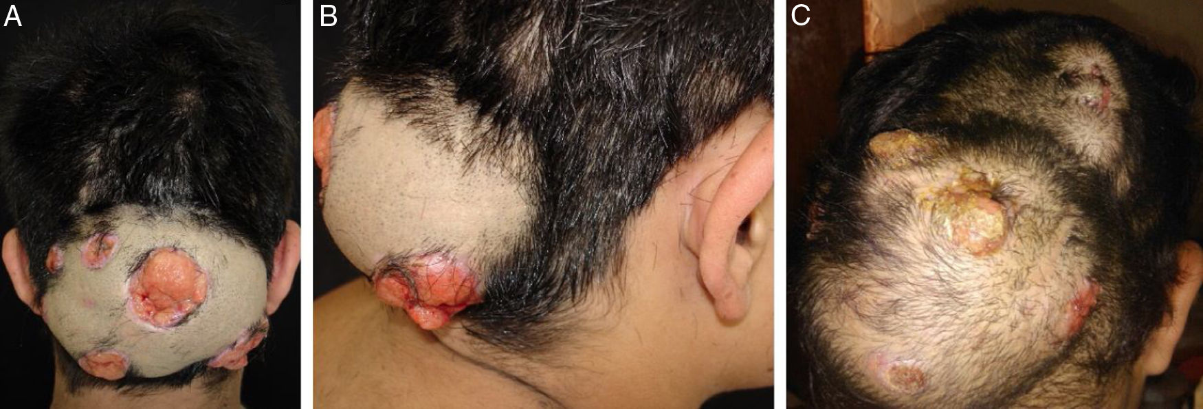
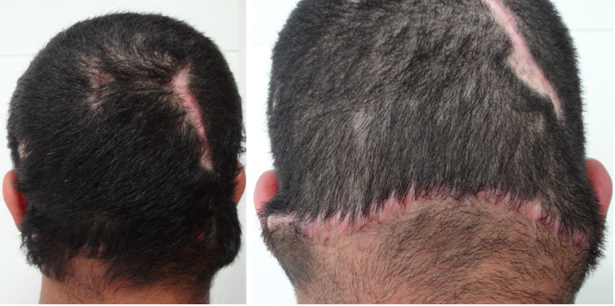
A 24-year-old immunocompetent male, with tumoral lesion in the occipital area which started at two years of age with areas of alopecia that progressively evolved to a tumor. His first consultation was at age 14, presenting at the clinical examination a hardened tumor with granular fundus ulcerations. He abandoned follow-up, returning ten years later with a considerable increase of the lesion (Fig. 1). Material analysis of ulcer scaling identified at direct microscope examination grains composed of septate hyaline hyphae, and micologial culture isolated Microsporum canis (Fig. 2). Histopathological examination revealed at the dermis and hypodermis clusters of septated hyaline hyphae of varied sizes involved by histiocytic Splendore-Hoeppli reaction with numerous multinucleated giant cells of foreign body type, besides neutrophilic exudate, edema and vascular congestion. No fistulated pathways were visualized promoting the continuity between the “grains” and the epidermal surface in the sample (Fig. 3). He denied use of any immunosuppressive medication, presented non-reactive serology for HIV, and had no other comorbidities. Associating the clinical aspect with the complementary tests, the diagnosis of pseudomycetoma by Microsporum canis was confirmed. The patient was submitted to surgical excision of the tumor and associated oral griseofulvin, one gram per day for two years. In a one year follow-up after the end of griseofulvin, the patient showed no signs of relapse (Fig. 4).

Figure 1.

Clinical aspect of pseudomycetoma at occipital lesion. A and B, patient with 14-years-old. C, patient with 24-years-old.

(0.17MB).
Figure 2.

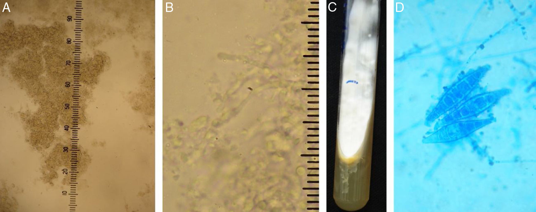
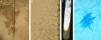
A and B, Direct mycological examination clarified with 20% KOH. ×100 augmentation, showing agglomerates of septated hyphae, and ×400 augmentation, identifying the structures of hyaline septated hyphae. C and D, fungal culture, with white filamentous colony and yellow pigmented agar and microculture with hyaline septated hyphae in the background and three macroconidia in the center (Cotton blue, ×400).

(0.13MB).
Figure 3.

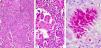
Histopathological examination. A, identifying grains surrounded by inflammatory suppurative process and lymphohistiocytic reaction (Hematoxylin & Eosin, ×100). B, grain composed of septated hyaline hyphae surrounded by eosinophilic material (Splendore-Hoeppli phenomenon) (Hematoxylin & eosin, ×200). C, evidencing the morphology of hyphae (PAS, ×200).

(0.34MB).
Figure 4.

Patient with clinical aspect after treatment.

(0.12MB).
Discussion

Chronic inflammatory and invasive forms of dermatophytosis are the result of an intense hypersensitivity reaction to the fungal infection, more frequent in immunocompromised individuals.1 The clinical presentations are kerion celsi, Majocchi granuloma and pseudomycetoma.2 Pseudomycetoma is an extremely rare mycosis, caused by the penetration of dermatophytes into the tissue from rupture of infected follicular epithelium. Ajello et al.3 reported several species of dermatophytes producing grains in tissues, including Microsporum canis, Trichophyton tonsurans and T. mentagrophytes. According to these authors, mycelium aggregates formed by the dermatophytes would be pseudo-granules and the term pseudomycetoma should be applied to this deep dermatophytic infection.4,5 The isolated agent in this case was compatible with the most frequent tinea capitis agent in Brazil.6,7

Clinical aspects of pseudomycetoma are identical to those of eumicetoma, yet in contrast to mycetomas, pseudomycetomas are more common in the scalp and do not have a history of trauma for its inoculation.8 Although the dermatophytic hyphae usually are more delicate than the eumicetoma agents at mycological examinations, the same does not happen in the clusters visualized on the histopathological examination.7–9 However, there is a difference between the two diseases at histopathology: mycetomas typically have sinus tracts through which fibrinopurulent exudate and grains are readily excreted; by contrast, pseudomycetomas lack sinus tracts.8 Therefore, isolation of the agent should be obtained with fungal culture, as here presented. Although more frequent in immunosuppressed patients, there are reports of cases in immunocompetent patients, and the eosinophilic reaction of Splendore-Hoeppli around pseudogranules is present in all cases of pseudomycetoma, highlighting the intense reaction of the organism against the fungus.4

The treatment of pseudomycetoma by M. canis is surgical excision of the fungal mass, since the systemic antifungal does not reach therapeutic concentrations,8 associated with oral griseofulvin1,6,7 until clinical and mycological cure.

Financial support

None declared.

Authors’ contributions

Ligia Rangel Barboza Ruiz: Approval of the final version of the manuscript; conception and planning of the study; elaboration and writing of the manuscript; obtaining, analysis, and interpretation of the data; effective participation in research orientation; intellectual participation in the propaedeutic and/or therapeutic conduct of the studied cases; critical review of the literature; critical review of the manuscript.

Clarisse Zaitz: Approval of the final version of the manuscript; conception and planning of the study; obtaining, analysis, and interpretation of the data; effective participation in research orientation; intellectual participation in the propaedeutic and/or therapeutic conduct of the studied cases.

Clarisse Zaitz: Intellectual participation in the propaedeutic and/or therapeutic conduct of the studied cases.

John Verrinder Veasey: Approval of the final version of the manuscript; elaboration and writing of the manuscript; critical review of the literature; critical review of the manuscript.

Conflicts of interest

None declared.

References
[1]
O. Barboza-Quintana, R. Garza-Guajardo, C. Assad-Morel, N. Méndez-Olvera.
Pseudomycetoma for Microsporum canis: report of a case diagnosed by fine needle aspiration biopsy.
Acta Cytol, 51 (2007), pp. 424-428
[2]
R. Isa-Isa, R. Arenas, M. Isa.
Inflammatory tinea capitis: kerion, dermatophytic granuloma, and mycetoma.
Clin Dermatol, 28 (2010), pp. 133-136
[3]
A. Ajello, W. Kaplan, F.W. Chandler.
Dermatophyte mycetoma: fact or fiction?.
PAHO Sci Pub, 396 (1980), pp. 135-140
[4]
M.A. Moraes, A.A. Machado, P. Medeiros Filho, C.M. Reis.
Dermatophytic pseudomycetoma: report of a case caused by Trichophyton tonsurans.
Rev Soc Bras Med Trop, 34 (2001), pp. 291-294
[5]
F. Botterel, S. Romand, M. Cornet, G. Recanati, B. Dupont, P. Bourée.
Dermatophyte pseudomycetoma of the scalp: case report and reviwe.
Br J Dermatol, 145 (2001), pp. 151-153
[6]
J.V. Veasey, B.A.F. Miguel, S.A.S. Mayor, C. Zaitz, L.H. Muramatu, J.A. Serrano.
Epidemiological profile of tinea capitis in São Paulo City.
An Bras Dermatol, 92 (2017), pp. 283-284
[7]
J.V. Veasey, G.S.C. Muzy.
Tinea capitis: correlation of clinical presentations to agents identified in mycological culture.
An Bras Dermatol, 93 (2018), pp. 465-466
[8]
E. Castro-Echeverry, K. Fiala, M.P. Fernandez.
Dermatophytic pseudomycetoma of the scalp.
Am J Dermatopathol, 39 (2017), pp. e23-e25
[9]
M. Tirado-González, E. Ball, A. Ruiz, Y. Rodriguez, C.E. Goudet, O. Finkel, et al.
Disseminated dermatophytic pseudomycetoma caused by Microsporum species.
Int J Dermatol, 51 (2012), pp. 1478-1482

How to cite this article: Ruiz LRB, Zaitz C, Lellis RF, Veasey JV. Pseudomycetoma of the scalp caused by Microsporum canis. An Bras Dermatol. 2020;95:372–5.

Study conducted at the Clínica de Dermatologia da Santa Casa de Misericórdia de São Paulo, São Paulo, SP, Brazil.

Copyright © 2020. Sociedade Brasileira de Dermatologia
Download PDF
Idiomas
Anais Brasileiros de Dermatologia
Article options
Tools
en pt
Cookies policy Política de cookies
To improve our services and products, we use "cookies" (own or third parties authorized) to show advertising related to client preferences through the analyses of navigation customer behavior. Continuing navigation will be considered as acceptance of this use. You can change the settings or obtain more information by clicking here. Utilizamos cookies próprios e de terceiros para melhorar nossos serviços e mostrar publicidade relacionada às suas preferências, analisando seus hábitos de navegação. Se continuar a navegar, consideramos que aceita o seu uso. Você pode alterar a configuração ou obter mais informações aqui.